 With the growth of immunotherapies, identifying biomarkers has become increasingly important. Traditionally, this is a process that is done individually for each marker of interest, however recently, multiplexing has allowed us to maximize the information we can glean from just one sample. Multiplexing can be used to identify multiple biomarkers on a single tissue sample, which helps provide additional information about the tumor microenvironment that can be used in the decision of whether a treatment is likely to be successful.
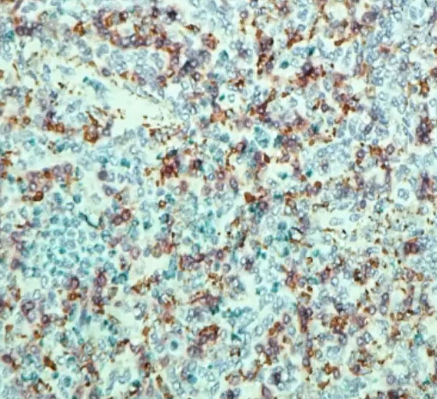
Multiplexing can either be done using fluorescent or chromogenic staining. In fluorescent detection, antibodies are conjugated with a fluorochrome and will emit light when stimulated with a light of a shorter wavelength. Alternatively, the antibodies can be conjugated with enzymes. The enzyme reacts with a substrate and leaves a chromogenic deposition at the site of reactivity.
Fluorescent vs Chromogenic Staining
Which method you select will depend on many factors, as each method has its advantages and disadvantages.
Overall, fluorescent staining lends itself better to multiplexing than chromogenic staining does. There are simply more colors and narrower emission spectra for each color with fluorescent staining, while chromogenic staining is confined to a few colors with a higher potential for spectral overlap when you try to use beyond 2 or 3. Therefore, the size of your panel is extremely important when selecting which method to use. Fluorescent detection offers the advantage of being able to better identify co-localized targets, whereas it can be difficult to distinguish single color from mixed color when targets are co-localized with chromogenic detection.
Advances in multispectral imaging technology have also made it easier to differentiate tissue autofluorescence from label fluorescence, as well as unmix different fluorophores that have overlapping excitation emission spectra for better quantification, making fluorescent staining even more advantageous.
So why would you use chromogenic detection? Well, a major drawback of fluorescence is it fades. If you need the stain to last, chromogenic staining is less susceptible to photobleaching. Chromogenic detection also has the advantage of greater sensitivity if you are using secondary antibodies for signal amplification.
Sequential vs Simultaneous Staining
Another decision to be made when developing a multiplexing procedure is whether to use sequential or simultaneous staining. This decision will largely be based off of the antibodies you are using, and the hosts they are from.
Sequential Staining: Sequential staining involves staining procedures done one after the other. So, the first primary antibody is applied with its detection system and chromogen. Then a second primary antibody is applied after the excess chromogen from the first staining has been removed. The benefit of this method is that it avoids cross-reactivity. Cross-reactivity is when an antibody has a high affinity for more than one antigen, so it reacts with an antigen besides the intended target. There are downsides to sequential staining though, such as, the possibility of incorrect double staining if you do not properly clear the sample of the leftover reagents from the first round of staining. It also cannot be used for co-localized targets, meaning targets in the same place. For example, it is fine if one stains the nucleus, another stains the membrane, another stains the cytoplasm, but you can’t do two nuclear markers, that stain the same nucleus.
Simultaneous Staining: In simultaneous staining, the antibodies can be applied simultaneously, which has the benefit of being less time consuming. Whether or not you can use this method however, depends on the antibodies you’re using. Not all antibodies can be used together at the same time. This only works if you use antibodies from two different hosts.
Want to learn more about multiplexing? Check out these great webinars, Multiplexing on Animal and Xenograft Tissue and IHC Multiplexing in Research and Clinical Application, available on elearn.nsh.org.
References:
https://www.biomol.com/resources/biomol-blog/overview-of-multiplex-immunohistochemistry
https://www.ptglab.com/news/blog/how-do-i-know-if-the-antibody-will-cross-react/
https://journals.sagepub.com/doi/full/10.1369/0022155417719419
https://www.nature.com/articles/s41374-020-0429-0
https://info.gbiosciences.com/blog/multiplex-immunohistochemistry
https://www.perkinelmer.com/CMSResources/Images/44-140165WHT_MultiplexingwithMSIfrommicetomicroscopy-9391.pdf
https://www.novusbio.com/ihc-detection
https://blog.cellsignal.com/pros-and-cons-of-different-multiplexing-techniques
#2021#IHCandMolecular#Blog