 The concept behind silver staining, the recognition that silver nitrate can stain organic substances, dates back to the 12th century, but it wasn’t until the mid-late 19th century that silver nitrate becomes more widely used in science. It is around this time that silver nitrate is utilized in photographic development and it slowly is tested for use in histological staining. By the 1970’s, Kerenyi and Gallyas, generally credited with the origin of the silver stain, begin using it from observation of proteins in gels. Camillo Golgi is generally credited with adapting silver staining for use in the central nervous system.
During silver staining, small, brown or gray-black metallic silver grains are deposited on the tissue structures. There are several ways this is accomplished, which is why there are several different classifications of silver staining methods; argentaffin methods, argyrophil methods, impregnation stains, silver oxidation-reduction stains, and metallic-metallic interactions.
Argentaffin reactions occur when ionic silver is reduced to metallic silver, depositing metallic silver at the reduction site. This type of silver staining is therefore used to identify molecules with strong reducing groups. For example, it is used in the detection of melanin, which has the ability to reduce silver. Fontana Masson is an example of such a silver stain used for melanin detection.
While argentaffin cells have the ability to be their own reducing agent, argyrophil cells do not. They absorb the silver ions but require the addition of a separate reducing agent in order for the silver to be converted into elemental silver. An example of this type of silver stain is Warthin-Starry, which is used in the detection of spirochetes.
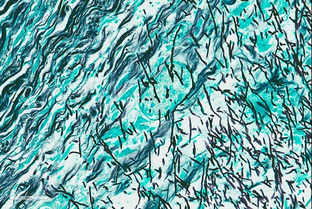
Impregnation silver stains also involve argentaffin reactions, but on the micro-level. In impregnation silver stains, such as the Bodian stain, argentaffin reactions are used to establish “silver seeds” and silver is absorbed onto these seeds. The Bodian stain is used on nerve fibers.
Silver oxidation-reduction stains are used to essentially artificially create reducing sites for argentaffin reactions. Oxidizing agents are applied, oxidation occurs, the extent of which is determined by the time and strength of the oxidation agent, and this causes the creation of reducing sites. This reaction is similar to what you would see in the PAS reaction, but the silver ion solution replaces the Schiff reagent. This is used in stains such as the Grocott modification of the GMS stain.
Lastly, we have metallic-metallic interactions with silver. This is primarily used to detect sites of antibody-antigen interaction. Tiny gold particles are used in place of a labeling enzyme. Because they are so tiny, electron microscopy is used to detect the gold particles. Metallic silver is deposited at the site of the gold particles to allow the sites to be seen with a light microscope.
References:
https://www.jstage.jst.go.jp/article/ahc1968/19/5/19_5_655/_pdf
https://www.tandfonline.com/doi/pdf/10.1179/2046023614Y.0000000054?needAccess=true
https://webpath.med.utah.edu/HISTHTML/MANUALS/GRIMEL.PDF
https://www.tandfonline.com/doi/pdf/10.1179/his.1996.19.3.183?needAccess=true
https://www.tandfonline.com/doi/full/10.1179/2046023614Y.0000000054?src=recsys
#2020
#Blog
#GeneralAnatomicPathology