Special stains are performed when there is something that is not adequately visualized with an H&E stain. They usually use dyes that are specifically good for whatever tissue component you are trying to highlight. One example of this is elastin. Elastin is a protein that makes up the elastic fibers found in connective tissue. Elastin is usually found in the skin and the walls of blood vessels and has the ability to resume its shape after stretching. Elastin, along with reticular fibers, another example of a connective tissue, are not easily distinguished with H&E and therefore require special stains. Pathologists may look at elastic fibers for diagnosis of Marfan Syndrome, emphysema, Giant Cell Arteritis, sun exposure, and more.
One of the stains most commonly used for elastic fibers is the Verhoeff’s Van Gieson, which is used for emphysema and vascular diseases. This stains the collagen red, while staining the elastic fibers black and the muscle yellow. The Verhoeff Hematoxylin used in this stain is only good for a few hours so it should be mixed right before use. To do this stain, overstain tissue in the Verhoeff hematoxylin for 10-60 minutes. Since it is a regressive stain you must differentiate using a 2% Ferric Chloride solution. Elastin has the strongest affinity to the Verhoeff Hematoxylin and will be differentiated last. It is then counter stained with Van Gieson. If you find your elastin to be pale, it could be because it is overdifferentiated, understained, you’ve used old Verhoeff Hematoxylin, or it spent too long in Van Gieson.
Aldehyde Fuchsin is another stain that is used for elastic. A 1950 article in the American Journal of Clinical Pathology by George Gomori describes this at the time, new, stain as an accidental product of experiments with Schiff’s reagent. He found that the stain would dye elastic fibers (both course and fine), as well as mast cells and beta cells in islets, a deep shade of purple. This stain may be used less often because it uses paraldehyde, which is a controlled substance and also begins to break down after first use.
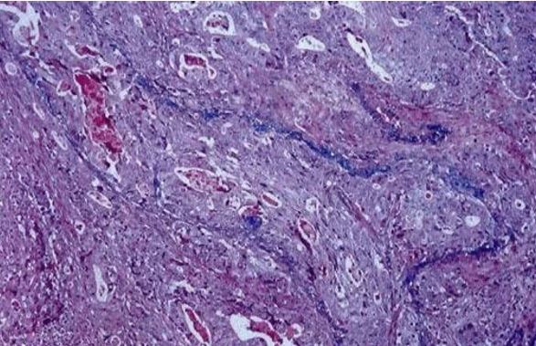
Victoria Blue is a dye that demonstrates elastic fibers in the background of H&E stains, dying them blue. This can be used for demonstrating venous invasion by tumor cells, particularly in the gastrointestinal tract, which is used during diagnosis of cancer. Victoria Blue H&E is used here instead of Van Gieson because it doesn’t require preparation of additional specimens.
VB-H&E Staining- Yutaka Tsutsumi, Noboru Onoda & R. Yoshiyuki Osamura (1990) Victoria Blue-Hematoxylin and Eosin Staining: A Useful Routine Stain for Demonstration of Venous Invasion by Cancer Cells, Journal of Histotechnology, 13:4, 271-274, DOI: 10.1179/his.1990.13.4.271
Orcein can also be used for elastic, provided that suitable dye batches are used. It will stain the elastic fibers brown. Orcein is also commonly used following Shikata’s modification, to demonstrate hepatitis B surface antigens.
References
https://academic.oup.com/ajcp/article-abstract/20/7_ts/665/4828701?redirectedFrom=fulltext
http://stainsfile.info/stain/elastic/methods-aldfuchsin.htm
https://www.tandfonline.com/doi/pdf/10.1179/his.1990.13.4.271?needAccess=true
http://education.med.nyu.edu/Histology/coursematerials/syllabus/stains.html
https://www.tandfonline.com/doi/abs/10.1080/10520290410001671335?src=recsys
#2020#Blog#GeneralAnatomicPathology